Chemistry prize

Born in 1960, he received his doctorate from Cornell University in 1988. He is currently the team leader of Howard Hughes Medical Research Institute.

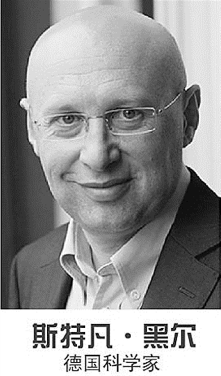
Born in 1962, he received his doctorate from Heidelberg University in Germany in 1990. At present, he is the director of Max Planck Institute of Biophysical Chemistry in Germany and the director of the branch of German Cancer Research Center.

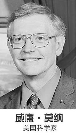
Born in 1953, he received his doctorate from Cornell University in 1982. At present, he is a professor of chemistry and professor of applied physics at Stanford University in the United States. Photo/Xinhua News Agency
Reasons for winning the prize
For a long time, scientists thought that optical microscope had a limit: it could not obtain a better resolution than half-light wavelength. With the help of fluorescent molecules, several winners of this year’s Nobel Prize in Chemistry skillfully bypassed this limit. Their breakthrough research has brought the optical microscope into the nanometer dimension. Today, nanotechnology is widely used in the world, and new knowledge is constantly produced, benefiting mankind.
Microscope has opened the door for people to observe the micro-world, but for a long time, people thought that optical microscope could not break through a limit: it could never get a higher resolution than the half wavelength of the light used-0.2 micron. However, three American and German scientists, Eric Bettzuege, William E. William E.Moerner and Stefan Hell, cleverly bypassed this limit.
Once the optical observation limit
For a long time, due to the limitation of optical properties, optical microscope can not obtain very accurate resolution. In 1873, ernst, a microscopist, deduced the "limit" that can be seen with an optical microscope according to the diffraction theory. That is to say, the distance between two points that can be optically resolved is always more than half the wavelength, and those targets whose diameter is less than 1/2 of the wavelength of visible light (the wavelength of visible light is between 0.4 micron and 0.7 micron) have closed the door to the world. For biologists, this also means that only the outline of mitochondria can be observed, and even smaller cell structures can not be observed, and it is even more extravagant to observe a single protein molecule. It’s like sitting on a plane and looking at the ground in a city. You can only observe large buildings, but you can’t see Mercedes-Benz cars and living residents. This famous law made scientists dare not expect to improve the resolution of optical microscope until the 20th century.
Find another way to bypass the limit
One morning in the early 1990s, Hale, who was lying in bed, suddenly had an inspiration. If a suitable laser is used to excite a fluorescent spot to emit light, and then a ring beam with a wavelength similar to the reflection halo is used to suppress the reflection, the observation scale of people will be greatly reduced. In the words of Tan Yanwen, a professor of physics at Fudan University, a doughnut is pressed on a big cake, and the central area of the doughnut is beyond the reach of traditional optical microscope.
Hale named this invention STED, that is, stimulated emission loss microscope, which was successfully introduced in 2000.
Eric and william moerner took a detour from another direction, which is different from Hale’s method of relying on quantum optics, which relies on a lot of calculations. Tan Yanwen told reporters that as early as 1995, Eric proposed to locate the cell molecule that people wanted to observe by calculating the center point of halo. The problem is that there are two molecules next to each other in the central area, and the positioning is easy to be "arrogant". This problem was not solved until the discovery of special fluorescent molecules in 2006. With the help of william moerner, Eric proposed the PALM model, that is, the same area was imaged many times, and only some molecules were made to fluoresce each time.
The two methods lead to the same goal, which makes the world that people can observe enter the nanometer scale. In theory, there is no structure that cannot be studied.
Basic invention rewrites biological cytology textbook
Tan Yanwen said that this year’s Nobel Prize in Chemistry and last year’s Nobel Prize in Chemistry have one thing in common, that is, the winning research is very basic. "Perhaps the fundamental reason for naming this research made by physicists as the Chemistry Prize is that its main application field is biology, and chemistry is closely related to biology." He quipped.
However, the emergence of high-resolution fluorescence microscope is enough to rewrite the textbook of cell biology, and the world of nano-sized object components is gradually enriched in front of people. Scientists can observe how molecules generate synapses between brain nerve cells; They can track the accumulation of related proteins in patients with Parkinson’s disease, Alzheimer’s disease and Huntington’s disease; They can also follow the changes of the internal protein of fertilized eggs when they differentiate into embryos.
However, at present, this technology is mostly used on fixed cells. Yu Yang, a staff member of the National protein Center, told reporters that this is because the imaging speed of the two methods is still relatively slow, and even the fixed cells may drift in different degrees, which affects their accuracy. It is worth noting that scientists are trying to reduce the influence of drift phenomenon through calculation and other methods, and ultra-high resolution imaging of living cells has also developed.
Nobel prize related to microscope
■ In 1925, the Nobel Prize in Chemistry was awarded to German colloidal chemist Richard Zsigmondy in recognition of his explanation of the multiphase nature of colloidal solutions and his breakthrough contribution in the field of colloidal chemistry. For this precise research, in 1903, he and Sidentov invented the ultramicroscope, and used ultramicroscope to observe and study colloids. We can observe the shape of any particle in one hundredth of a meter, and see the fine structure of organisms or the existence of colloidal particles.
■ In 1953, the Nobel Prize in Physics was awarded to Zelnik in university of groningen, the Netherlands, for proposing the phase contrast method, especially inventing the phase contrast microscope. Phase contrast microscope is a special microscope, especially suitable for observing objects with high transparency, such as biological slices, oil films and phase gratings.
In 1982, the Nobel Prize in Chemistry was awarded to Aaron Klug, a British chemist, who used high-resolution electron microscopy and X-ray diffraction analysis techniques to conduct in-depth research on the complex system of nucleic acids and proteins. He studied the structure of many viruses, put forward the structural theory of spherical virus, and found out the two components of protein and nucleic acid in tobacco mosaic virus (TMV), a coryneform virus, and won the Nobel Prize.
■ In 1986, half of the Nobel Prize in Physics was awarded to Ernst ruska of the Fritz-Haber Institute in Berlin, Germany, in recognition of his basic work in the field of electro-optics and the design of the first electron microscope; The other half was awarded to Buennig, a German physicist, and rohrer, a Swiss physicist at IBM Zurich Research Laboratory in Ruxilikang, Switzerland, for designing the scanning tunneling microscope.